|
Gram-staining Procedure
Gram-staining is a four part procedure which uses certain dyes to make a
bacterial cell stand out against against its background. The specimen should be
mounted and fixed on a slide before you procede to stain it. The reagents you
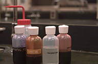
will need to successfully perform this operation are:
- Crystal Violet (the Primary Stain)
- Iodine Solution (the Mordant)
- Decolorizer (ethanol is a good choice)
- Safranin (the Counterstain)
- Water (preferably in a squirt bottle)
Before starting, make sure that all reagents, as well as the squirt-bottle of
water, are easily accessible because you won't have time to go get them during
the staining procedure. Also, make sure you are doing this near a sink because
it can get really messy. Wear a lab coat.
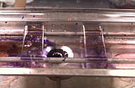
STEP 1: Place your slide on a slide holder or a rack. Flood (cover
completely) the entire slide with crystal violet. Let the crystal violet stand
for about 60 seconds. When the time has elapsed, wash your slide for 5 seconds
with water. The specimen should appear blue-violet when observed with the naked
eye.
STEP 2: Now, flood your slide with the iodine solution. Let it stand about a
minute as well. When time has expired, rinse the slide with water for 5 seconds
and immediately procede to step three. At this point, the specimen should still
be blue-violet.
STEP 3: This step involves addition of the decolorizer, ethanol. Step 3 is
somewhat subjective because using too much decolorizer could result in a false
Gram (-) result. Likewise, not using enough decolorizer may yield a false Gram
(+) results. To be safe, add the ethanol dropwise until the blue-violet color is
no longer emitted from your specimen. As in the previous steps, rinse with the
water for 5 seconds.
STEP 4: The final step involves applying the counterstain, safranin. Flood
the slide with the dye as you did in steps 1 and 2. Let this stand for about a
minute to allow the bacteria to incorporate the saffranin. Gram positive cells
will incorporate little or no counterstain and will remain blue-violet in
appearance. Gram negative bacteria, however, take on a pink color and are easily
distinguishable from the Gram positives. Again, rinse with water for 5 seconds
to remove any excess of dye.
After you have completed steps 1 through 4, you should blot the slide gently
with bibulous paper or allow it to air dry before viewing it under the
microscope. DO NOT RUB THE SMEAR!
|